Growing an embryo against the record
The embryo divides, folds and lays out its organs stage by stage, and on 18 September 2026 it reached hatching for the first time. We timed every stage from published observations, checked each against them, and reconciled seven sources that disagreed.
What the genes are doing, hour by hour — sixty single embryos, one an hour across four days, 9,069 genes measured. Scrub through development and watch each one switch on, including the moment condensin II shuts down for good.
What switches on when — the same embryos as a network: 409 genes placed on a time axis by when they peak, joined to the genes whose profiles follow theirs. The chromosome machinery goes dark, a knot of patterning factors fires in twelve hours, and the synaptic genes climb for the rest of development.
Cell cycles from the lineage, not the hour
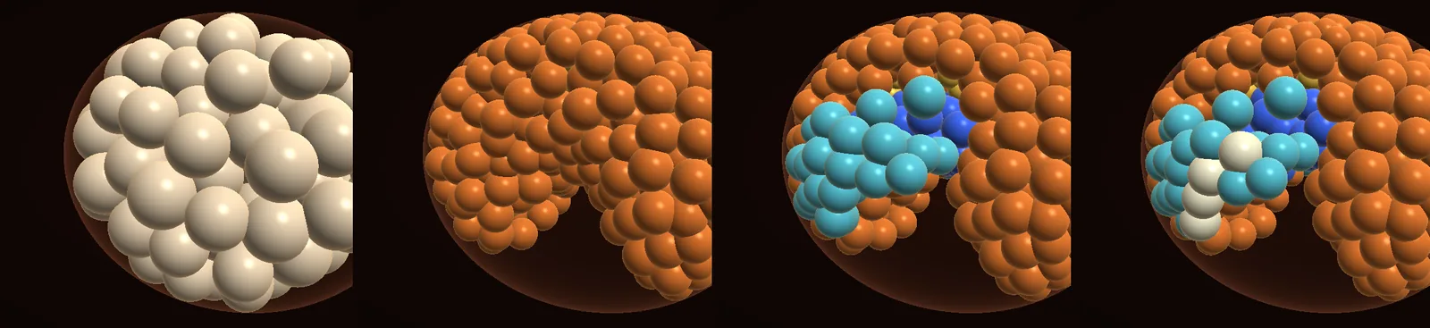
The embryo's early cells now divide on the measured clock: 45, 44, 44, 54, 72 and 111 minutes for the first six generations, faster than the "approximately once per hour" of the first staging series (Gabriel et al. 2007), and lengthening as the cells shrink. The times come from Heikes et al. 2023, who timed every cell in nine embryos and shared the table. Every one of the 22 cells the simulation shares by name with the lineage falls within the measured spread between embryos. That match is a fit, since it came from the same table. The real test was that everything downstream still held: the germ cells still go inside at about 8 hours and end in the third trunk segment, and the epithelium forms on time with about 480 cells, closer to the ~500 observed than before.
How this is built
- Data
- Heikes et al. 2023's lineage, every cell timed in nine embryos, from the table they shared on GitHub. Stage times and cell counts from Gabriel et al. 2007.
- Built with
- Our embryo simulation, one web page of soft-sphere cells, takes the first six generations' cycle lengths from the lineage's means. The spread between cells follows Schnabel 1997's measurements in the worm C. elegans. The chart plots those means against the earlier hourly rule.
- Checked
- Division times of 22 named cells against the lineage: all within the measured spread, but that is a calibration, not a test. Against Gabriel's stages, independently: partial. The 4- and 60-cell stages fall in their bands, the 16- and 32-cell stages up to 0.3 h early, and the epithelium forms at 15 hours with 470–482 cells against ~500.
- Limits
- On 19 September the late divisions changed to follow the expression data: one slow round after round 8, and none after 48 hours. The hatching count above predates that change and has not been retested.
- More
- How the whole project is built · every rule and test
Seven sources, one timeline
We lined up every published timing of the same embryo: the 2007 series, and four groups since (Game & Smith, Mapalo, Smith and Treffkorn). Almost every stage agrees within two hours, except one. The 2007 series puts limb buds at ~26 hours, while everyone after puts them at 30–35, and Game & Smith say outright that the original is about 10 hours early. Because the stages either side agree, this is a local error, not a warmer or cooler laboratory. The simulation now grows limb buds from 30 hours to full size at 45, pharynx at 31, ganglia at 40, claws at 52, and hatches at 100 hours.
How this is built
- Data
- Every staging and timing we have read for this embryo: Gabriel et al. 2007, Heikes et al. 2023, Game & Smith 2020 and 2025, Mapalo et al. 2024, Smith et al. 2016, Treffkorn et al. 2022 and Gross & Mayer 2015.
- Built with
- We lined them up stage by stage in one table and chose a reconciled time for each stage. The simulation's stage rules (limb buds, pharynx, ganglia, claws, hatching) follow that column.
- Checked
- A full run against all 19 stages: partial. Every stage is reached on the reconciled times in two runs, with 1,086–1,121 cells at hatching against 1,140 ± 60. Limb buds at full size are 15–16 µm long against ~18 (close).
- Limits
- Only Game & Smith (23 °C) and Levin (20 °C) state a temperature; for "room temperature" we assume about 21 °C (19–24). From elongation on, stage shapes and times are prescribed, not emergent.
- More
- How the whole project is built
A dataset's clock, set right
We set right the clock of a widely reused dataset: single embryos sequenced through development by Levin et al. 2016. Its figure and supplement say the series spans 49.5 hours, while its methods and database labels say about 98. We compared each of their 62 embryos, gene by gene, with Yoshida et al. 2017's daily egg samples. On the ~98-hour axis the embryos fall onto egg days 1 to 5 in order, with hatching at the end. On the 49.5-hour axis, embryos at 23–32 hours would look like three-day-old eggs, which is impossible. So the supplement's minutes are halved. The corrected axis places a batch of nine unusual embryos exactly where the series changes most, which needs a closer look.
How this is built
- Data
- Levin et al. 2016's 62 single-embryo transcriptomes and Yoshida et al. 2017's daily egg samples (CC BY 4.0), both public at NCBI.
- Built with
- We aligned Levin's 12,734 transcripts to the nHd_3.1 genome with minimap2 (
layer2.py): 12,724 placed, on 9,069 genes. Then we matched each embryo to the egg day it most resembles, over 2,919 variable genes scaled within each dataset. The chart plots that match. - Checked
- The two datasets agree gene by gene: Spearman's rho 0.52 across 8,541 genes, against 0.008 with the gene labels shuffled (pass). On the ~98-hour axis the embryos fall onto egg days 1 to 5 in order.
- Limits
- Levin's embryos grew at 20 °C and Yoshida's eggs at 18 °C; we assume that gap doesn't shift the match. One embryo per time point, with no replicates, and no embryos sampled between about 20 and 32 hours.
- More
- Genomics · how the whole project is built
Organs, and a gradient nobody put there
From 40 hours, four ganglia gather under the belly in front of each leg pair, the fourth a few hours later, as in Martin et al. 2017's drawing of tardigrade neural development. No cell count was set. Yet the ganglia come out largest at the front and smallest at the back (on average 42, 27, 26 and 20 cells), close to the pattern Zantke et al. counted in adult Macrobiotus (ganglia 2–3 about 28% smaller than ganglion 1, ganglion 4 about 55% smaller). It follows from the curled body: the front of the trunk carries more surface cells. It's suggestive, not proof: a different species, and adult nuclei rather than embryonic cells.
How this is built
- Data
- Ganglion placement and order from Martin et al. 2017's drawing of neural development; ganglia at 40 hours from the reconciled timeline; adult nuclei counts in Macrobiotus hufelandi from Zantke et al. 2008, for comparison only.
- Built with
- A rule in the simulation: belly surface cells within 10 µm of a point just in front of each leg pair gather beneath the skin over 4 hours, ganglia 1–3 at 40 hours and ganglion 4 at 44. No cell count is set.
- Checked
- Over five runs the ganglia hold 42, 27, 26 and 20 cells, against Zantke's adult gradient: close, and emergent rather than fitted.
- Limits
- Where the ganglia form is prescribed, and ganglion 4's delay is our assumption. A different species, adult nuclei rather than embryonic cells, and simulated cells about three times the diameter of neuronal nuclei.
- More
- The brain and nerve cord · how the whole project is built
Cells that fill the egg
A second embryo takes a different approach: every cell owns a share of the egg and the shares fill it exactly, so cells can neither overlap nor leave gaps. It runs to about a thousand cells, elongates into the comma shape, opens a gut lumen and carries signals across the faces cells share — and the same program runs a nematode worm, whose embryos have been measured cell by cell. See it, and the worm check.
We also wrote down what the model predicts the genes should do, before we looked. Eight predictions with hours attached, committed, then tested against 62 single embryos: two held, five failed, and the biggest failure rebuilt the model’s whole second half. Every prediction drawn over what the genes actually did.
Cells with membranes
The first sixteen hours now also run with a real membrane around every cell, computed on a graphics card: turn the embryo in 3D as it packs itself from one cell to 485 and gastrulates — cells leaving the surface and moving inside, the four germ cells set aside, and the surface closing into an epithelium. It is shown with its known fault: cells overlap as the embryo crowds, and we say why on the page.
Where you put the dot matters. Models like these need a starting point for each cell, and in the worm — where tracing gives you nuclei and not cell outlines — that point is usually the nucleus. We measured how far off that is: across 60,701 cells, a nucleus sits about a quarter of a cell radius from its cell's true centre, always leaning the same way. What that costs, and why the two obvious fixes both fail.
What's still drawn rather than grown
- From elongation on, shapes are prescribed targets: the comma, pouches, buds, pharynx, ganglia and the roll in the shell. Replacing them with tissue mechanics is the next big step.
- Limb buds don't yet separate into lobes. Our cells are spheres about 5–6 µm across, while real epidermal cells are columnar, about 3 µm wide, and four lobes won't fit at sphere size.
- The four-cell embryo is flat in one mechanics rule and twisted in another. Nobody has published which is right; it's on our list to film.
Every rule, its source and its test are in the project's graph. The build log has the day's work.
Embryos, from the papers


